top of page
Tour our Technology

Optomap Monaco by Optos
This one-of-a-kind instrument combines Optomap ultra-widefield photography technology with autofluorescence and SD-OCT creating a fast, convenient retinal imaging tool, all in as little as 90 seconds. Our doctors recommend an Optomap screening at every comprehensive eye examination to help detect and manage eye diseases.

Nidek Auto-Refractor and Keratometery
This device helps measure the approximate starting point of a patient’s prescription as well as measure the curvature of the front of the eyes.

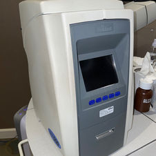
Nidek Non-Contact Tonometer
This instrument utilizes a light puff of air to determine the intraocular eye pressure.

Nidek Lensometer
This instrument is utilized by our technicians and opticians to measure the prescription on patients’ glasses. Patients are asked to bring their current eyewear to their comprehensive exam. This helps our doctors understand what our patients are accustomed to wearing.

Zeiss Humphrey FDT Visual Field
This device is used to screen for peripheral vision defects. (Please note that the office is not currently using this device during the pandemic.)

Digital Exam Lane
Welcome to your Digital Exam Lane! Claremont Optometry is proud to use the Nidek digital refraction lane system which consists of the Digital Control Panel and Phoropter as well as an Electric Chair and Topcon Slit Lamp. These instruments help make for a more comfortable, precise and efficient exam.

Nidek Digital Control Panel
Our doctors utilize this control panel during the examination to determine a patient’s refractive status at distance and near, and diagnose binocular abnormalities.

Zeiss Humphrey Visual Field
This instrument utilizes tiny points of light in a controlled environment to create a map of a patient’s vision. The results are interpreted by our doctors to determine if a patient has any loss of peripheral or central vision due to various ocular conditions (e.g.: glaucoma, macular toxicity due to certain medications, etc.) as well as neurological conditions that can affect vision (e.g.: brain tumors, stroke, etc.).

Optovue Optical Coherence Tomography (OCT) with anterior and posterior capability
This is a noninvasive imaging device that utilities light to capture a highly detailed image of various ocular structures including the macula, retina, optic nerve and cornea. These specialized images are used to manage various ocular conditions such as glaucoma, macular degeneration, epiretinal membrane, central serous choroidopathy, papilledema, keratoconus, and many more.

Nidek Corneal Topographer
The corneal topographer is a noninvasive imaging technique used to map the surface of the cornea, or front of the eye. The result is a 3D map that is interpreted by the optometrist to determine if the patient has any number of corneal conditions, e.g. keratoconus, pellucid marginal degeneration, pterygium, corneal scarring, post-LASIK surgery, etc.

PacScan Plus A-Scan
The A-scan is a highly specialized device used by select doctors to very precisely measure the length of the eyeball. Today, we use it to calcuate the intraocular lens power for infant cataract surgeries and also to monitor myopia (nearsightedness) progression.

Eaglet Eye Surface Profiler
Creates a 3D map of the cornea, limbus and sclera, assisting in a highly customized scleral lens.
bottom of page


